- PATIENT FORMS | REQUEST A CONSULTATION | CONTACT US
- 1-844-NSPC-DOC
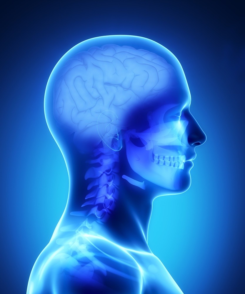
Stereotactic Radiosurgery for Arteriovenous Malformation (AVM)
Cerebral arteriovenous malformations (AVMs) are irregular networks of veins and arteries in the brain. Stereotactic radiosurgery is one method of treatment to avoid or treat an AVM rupture, which can cause blood to leak in the brain and may lead to a hemorrhagic stroke. AVMs in the brain can also reroute blood from the necessary tissue, which may cause neurological damage. Blood that pools in the brain may also compress healthy brain tissue, which can lead to seizures.
Although AVMs are generally congenital and present at birth, they often are not discovered for many years. An arteriovenous malformation can also develop in the spine or other areas in the body.
What Are the Treatments for Cerebral Arteriovenous Malformations?
Brain arteriovenous malformations have three types of treatment methods.
Embolization is an endovascular technique that involves occluding (blocking) some or all of the AVM with a glue-like material. Embolization may be used as part of a preoperative procedure to help make the surgical removal of the AVM safer by reducing the potential blood loss during surgery. Embolization may also be used to shrink the AVM so that it is smaller and better able to be treated with stereotactic radiosurgery.
Surgical removal of the AVM employs microneurosurgical techniques to resect the AVM. Once removed, the AVM does not return. However, whether your AVM is a good candidate for microsurgical resection depends on a number of factors including the size, the location, and the site of the venous drainage.
Radiosurgery for AVM is generally a one-time treatment for smaller AVMs, but may require several sessions for larger AVMs. This alternative to invasive surgery uses x-ray or gamma-ray beams with pinpoint accuracy to target the malformation. Results are not immediate and can take up to 2 or 3 years to see complete occlusion of the vessels. During this time, the AVM may still be at risk for bleeding.
Radiosurgery is often a one-time treatment that shrinks the AVM by constricting the blood flow to the malformation.
- 3-D imaging for precise targeting of the tumor or lesion, both in preparation for the radiosurgery and for positioning during the session
- Immobilization of the patient to keep the targeting precise.
- Gamma-ray or x-ray beams with pinpoint accuracy.
Your specific AVM treatment plan may involve a blend of these techniques.

NSPC’s expert physicians are here in our Long Island, NY centers to provide leading-edge radiosurgical options for AVMs and other brain and spine conditions. Make an appointment with a specialist at one of our New York locations—and find out if this state-of-the-art treatment can help you.
Connect With Our 7 Convenient Locations
across Long Island, NY
Our expert physicians, surgeons and doctors are ready to serve you at our 7 convenient locations across Long Island, NY. Connect today to learn how our award winning, world class experts can help.
4250 Hempstead Turnpike Suite 4,
Bethpage, NY 11714
(516) 605-2720
COMMACK
353 Veterans Memorial Hwy,
Commack, NY 11725
(631) 864-3900
One Hollow Lane, Suite 212
Lake Success, NY 11042
(516) 442-2250
MANHATTAN
215 E. 77th Street Ground Floor
New York, NY 10075
(646) 809-4719
PORT JEFFERSON STATION
1500-8A Route 112,
Port Jefferson Station, NY 11776
(631) 828-3001
100 Merrick Road, Suite 128W
Rockville Centre, NY 11570
(516) 255-9031
WEST ISLIP
500 Montauk Hwy
West Islip, NY 11795
(631) 983-8400
World
Class
Expertise
For over 50 years & 350,000 patients NSPC has been a trusted global medical leader.
Contact us today for an appointment or consultation.