- PATIENT FORMS | REQUEST A CONSULTATION | CONTACT US
- 1-844-NSPC-DOC
A Good Solution For Patients With Osteoporosis Who Need Surgery For A “Slipped Disc” or Spondylolisthesis

Tale of Two Pathologies Contributing to Lumbar Stenosis with Spondylolisthesis
April 28, 2023NSPC Physicians Collaborate on Ebook: Collection of Real-Life Clinical Cases
July 26, 2023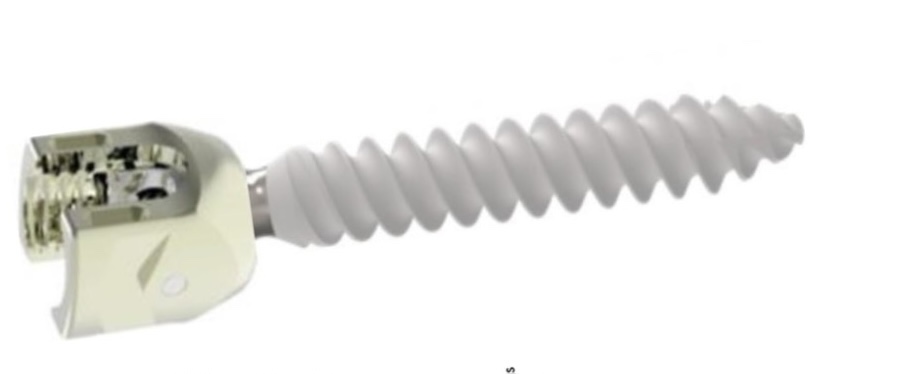
hydroxyapatite coated pedicle screw
I specialize in and am very familiar with patients who have osteoporosis and require spinal surgery for degenerative lumbar disease. I am particularly interested in treating those osteoporotic patients with spinal stenosis and a concurrent spondylolisthesis or “slipped spine” or “slipped disc”. It is actually not the disc that slips in a spondylolisthesis, but one vertebra slips forward on the one below. As a result of this degenerative laxity the spine is signaled to make arthritis such as enlarged joints or thickened ligaments to compensate for the chronic instability that mainly contribute to narrowing of the spinal canal where the nerves travel. This is called spinal stenosis. Patients often see their MRI reports and note that they often have been described as having this aforementioned condition. I believe that perimenopausal, menopausal, and postmenopausal women (40-60 years of age) would benefit greatly because of their degree of activity and expectations from a new technology that my spine team is offering. However, this is not to say that men or women at any age who do require surgery would not benefit. A patient comes into the office and requires surgery to fix their slipped disc or spine and they say to me, “doctor but I have osteoporosis.” I tell them that we have available special coated screws that are designed for patients with osteoporosis that will do a much better job holding their bones together.
(Fig 1) Hydroxyapatite Coated Pedicle Screw
Pedicle screws can be coated with a thin layer of hydroxyapatite (Fig. 1), which is a natural form of calcium apatite present in human bone and plays a role in the structural strength of bone and in bone regeneration. By coating the screws with hydroxyapatite this can improve screw fixation or bonding at the bone-implant interface
Here is a case in which we felt it was important to use this type of screw in a young female patient with severe osteoporosis with lumbar stenosis and a “slipped disc”:
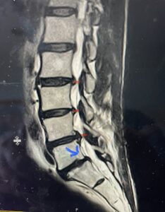
(Fig. 2) T2-weighted lumbar MRI demonstrating severe lumbar stenosis (red markers) and a grade 1 spondylolisthesis at L5-S1 (blue arrow)
This 59 year-old female had severe bilateral leg pain and numbness over a 6-month period. The patient had failed conservative treatment with physical therapy, chiropractic care, and medications. MRI revealed that she had severe lumbar stenosis with a grade 1 spondylolisthesis or “slipped disc” at L5-S1 (Fig. 2). In addition, she had previously undergone both front and back surgery for severe cervical stenosis where her posterior hardware had failed because of her severe osteoporosis requiring us to remove the posterior hardware. This required her to have an anterior or front operation which allowed better fixation to her spine because of the load-sharing nature of the interbody grafts in addition to her anterior cervical plate (Fig. 3).
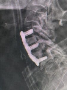
(Fig. 3) lateral intraoperative cervical x-ray demonstrating good alignment after C4-C6 anterior cervical discectomy and interbody fusion with plate. Note the interbody grafts help load-share the plate in this patient with severe osteoporosis
Because she had failed conservative management of her lumbar stenosis and spondylolisthesis it was decided to perform a lumbar laminectomy and fusion of the L5-S1 segment. Because of her history of severe osteoporosis and hardware cut-out from the bone, we decided to offer her hydroxyapatite-coated screws to provide fixation of her L5-S1 segment (Fig. 4a and b)
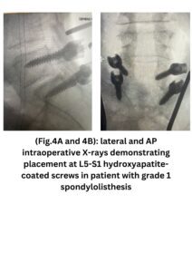 Intraoperatively, we noticed that with the aid of these special screws we got good fixation with the hope that over the next 6 months there would be excellent osteointegration of the screws because of the coating of hydroxyapatite. She had an uneventful postoperative course with improvement of her leg symptoms.


