- PATIENT FORMS | REQUEST A CONSULTATION | CONTACT US
- 1-844-NSPC-DOC
Awake Craniotomy Improves Outlook For Brain Tumor Patients Dr. Rak Performs Procedure

Increasing Awareness of Spina Bifida
July 10, 2015Grand Rounds 8/21/2015
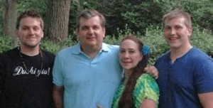
July 23, 2015HIGH SCHOOL sweethearts Donald and Melanie Squire of East Northport had just returned home from a relaxing vacation in Aruba. But the couple became concerned when Donald’s eye began to twitch uncontrollably and he experienced a loss of peripheral vision. Suspecting a possible stroke, Melanie, an ICU nurse at Huntington Hospital, called 9-1-1. Donald was taken by ambulance to Huntington Hospital where a number of tests revealed that he was suffering from glioblastoma, the most aggressive type of brain cancer. His tumor was in the right occipital lobe, in the back of the brain.

A team of specialists led by neurosurgeon Ramin Rak, MD, and included neuro-oncologist Paul Duic, MD, determined that Donald was an excellent candidate for awake craniotomy. This is a procedure in which the tumor is removed surgically while the patient remains awake in order to prevent damage and protect eloquent parts of the brain such as speech, movement and vision, with a process called direct brain mapping. “In the past, tumors similar to Mr. Squire’s in the eloquent brain, or dominant hemisphere of the brain, would have been considered inoperable or very risky,” explained Dr. Rak
“By monitoring the patient’s sensory and motor capabilities during surgery, such as vision, language and movement of the extremities, we minimize risk. We are also now able to map and monitor not only gray matter, but also white matter tracts during awake brain surgery.”
Director of Huntington Hospital’s Brain Tumor Program and one of the most experienced neurosurgeons performing this procedure in the New York metropolitan area, Dr. Rak is a board-certified neurosurgeon with extensive experience in awake craniotomies and micro-neurosurgical techniques. He is also an expert in neuronavigation in skull-based surgery as well as brain mapping. His extensive education includes clinical neurosurgery fellowships with both the National Institutes of Health (NIH) and the North Shore-LIJ Health System.
Improved technologies and newer types of anesthesia make the procedure more precise, safe and easier to conduct than it was just a few years ago. Functional magnetic resonance imaging (fMRI) is used during the surgery to measure brain activity by detecting associated changes in blood flow. Advanced micro-neurosurgical techniques allow Dr. Rak to remove more of the tumor and less healthy tissue, with greater accuracy. Patients feel no pain during the surgery, despite their awake state. This is because the brain, when touched directly, does not feel pain, and because local and regional anesthesias are used on the surrounding tissues such as the scalp and skull.
Another integral member of the surgical team is neuropsychologist Gad Klein, Ph.D., who works closely with Dr. Rak on awake craniotomies and performs testing both before and during the procedure. Dr. Klein asked Donald to perform a number of activities while Dr. Rak stimulated the relevant areas of the brain in order to prevent damage to vital functions. Other members of the team included an anesthesiologist who carefully adjusted anesthesia levels throughout the procedure’s multiple phases and a neurophysiologist who monitored the brain’s electrical stimulations.
Because of the aggressive nature of the tumor, Donald required additional surgery five months after the initial procedure. But because of Dr. Rak’s expertise in brain mapping technology, Donald did not need to be awake during the second surgery. Information stored on a computer from the first surgery, including microscopic images, served as a map for Dr. Rak during the second surgery.
Donald was able to go home just two days after the surgery. He is now receiving rounds of radiation and chemotherapy, standard post-surgical treatment for glioblastoma.
“I feel great,” he said recently while expressing his gratitude to the entire team. “I’m so thankful that my wife knew Dr. Rak. And I just try to be thankful for every moment I have with my wife and sons.”
