- PATIENT FORMS | REQUEST A CONSULTATION | CONTACT US
- 1-844-NSPC-DOC
Neurosurgeons Collaborate to Treat Giant Symptomatic Meningioma

A Good Solution For Patients With Osteoporosis Who Need Surgery For A “Slipped Disc” or Spondylolisthesis
July 26, 2023Neurosurgeons Collaborate to Treat Giant Symptomatic Meningioma
CLINICAL CASE:
 For several days, an otherwise healthy, 62-year-old woman displayed confusion, headaches, and word-finding difficulties. She was brought to the emergency department at a local hospital and underwent a CAT scan of the head which showed a large mass on the left frontal region with mass effect, consistent with a brain tumor.
The patient was admitted to the hospital’s intensive care unit and started on steroids and seizure prophylaxis medications. A subsequent MRI scan, with and without contrast, revealed that the large mass was a giant left frontal extra-axial meningioma (Fig. 1 right).
CLINICAL MANAGEMENT AND TREATMENT:
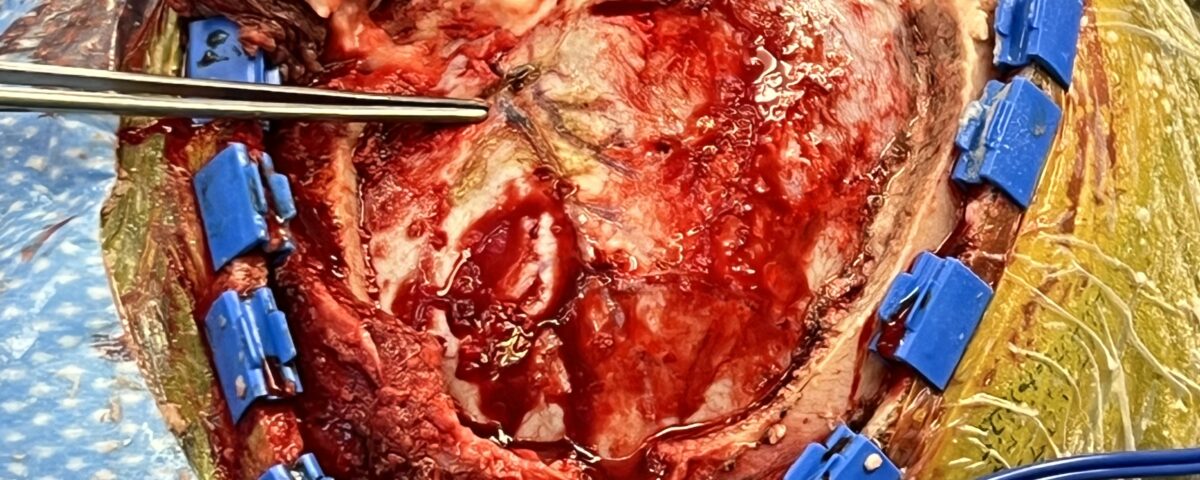
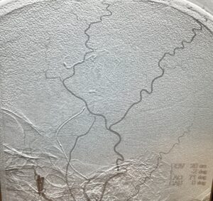
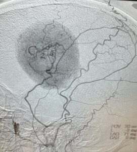
The case was discussed in detail with the patient’s family and the patient herself, whose speech had improved somewhat due to the steroids. Considering the size and the location of this tumor, neurosurgeon Ramin Rak M.D. recommended resection of the tumor with the aid of neuronavigation. To reduce the size of the tumor prior to the craniotomy, Dr. Rak enlisted his colleague, cerebrovascular neurosurgeon Jonathan Brisman M.D., to embolize some of blood vessels feeding the tumor (Fig. 2 above).
Successful embolization of the feeding vessels from the middle meningeal artery significantly helped the tumor resection that followed 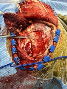 (Fig. 3 right).
Surgery was performed by Dr. Rak. A craniotomy exposed the underlying tumor which was identified and removed using microsurgical and fine surgical techniques.
A piece of DuraGen was then trimmed properly and placed over the cortex and the surrounding dura. The craniotomy bone flap was put back together using a mixture of round titanium bur hole covers and one straight plate. There were no complications, and the patient was transferred back to the intensive care unit in stable condition. Since her discharge, she has done very well and has made a full recovery.
Neurosurgeons Collaborate to Treat Giant Symptomatic Meningioma

Authors
To learn more about the author, click his name or photo:

