- PATIENT FORMS | REQUEST A CONSULTATION | CONTACT US
- 1-844-NSPC-DOC
39 year old physician with rhinorrhea and an encephalocele
Carotid Cavernous Fistula (CCF) Study
October 27, 2021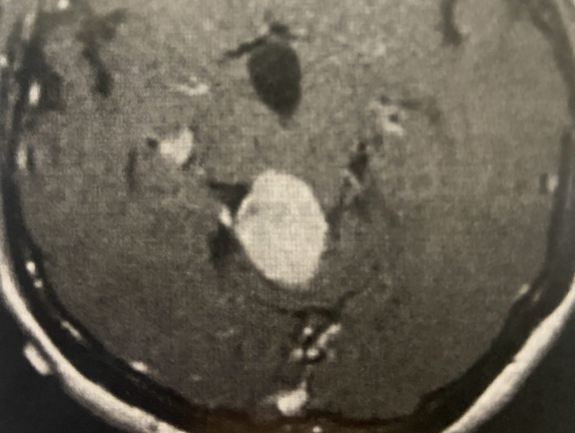
Pineal Region Meningioma
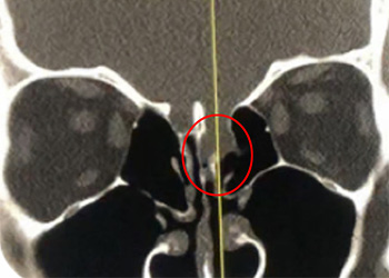
February 2, 2022This is a 39 year old physician with a long history of sinus issues, who more recently had noted an increased drainage of clear fluid from his nose. Testing of the fluid for Beta-2-transferrin demonstrated that it was consistent with cerebrospinal fluid (csf). MRI and CT scan demonstrated an encephalocele in the frontal most part of the anterior skull base, on the left, just off midline (Figures 1a,1b,1c). For the last 3 weeks, he has also been having headaches and neck pain.

Figure 1a – Coronal CT bone window

Figure 1b – Sagittal CT bone window

Figure 1c – Sagittal T2 MRI
The patient was sent to the hospital. He was noted to be afebrile with a normal white blood cell count. Nonetheless, to be safe, he was started on broad spectrum antibiotics. Two days later he was brought to the operating room and underwent a left frontal craniotomy to repair the encephalocele. Brainlab neuronavigation was used to avoid violating the frontal sinus, and a lumber drain was used to give adequate brain relaxation. A sizeable defect was noted in the skull base. Brain tissue was amputated from beneath the defect, and the defect was repaired with allograft.
The CSF never grew out any organisms and seemed otherwise unremarkable. The lumbar drain was left in place for 3 days postoperatively. The patient had no more csf rhinorrhea or headaches, and was discharged neurologically intact.
Encephaloceles are defects in which the brain exits the skull through defects in the skull. They are usually discovered at birth, and usually present as obvious protrusions from the head. However, it is possible to have an encephalocele present during adulthood, as in this case. The skull defect through which the brain protruded was from the anterior skull base into the ethmoid sinus, so it would not have been grossly noticeable. It is unclear whether some of this patient’s chronic sinus issues may have been secondary to this encephalocele, or may have ultimately led it to tear, and start leaking csf from the nose. Regardless, in light of the csf rhinorrhea, surgery was indicated to repair the defect to stop the csf leak and prevent the development of meningitis or brain abscess.
CATEGORY: BRAIN // 39 YEAR OLD PHYSICIAN WITH RHINORRHEA AND AN ENCEPHALOCELE
39 year old physician with rhinorrhea and an encephalocele