- PATIENT FORMS | REQUEST A CONSULTATION | CONTACT US
- 1-844-NSPC-DOC
Atypical Convexity Meningioma
Man in 40’s with progressive confusion and headaches
October 27, 2021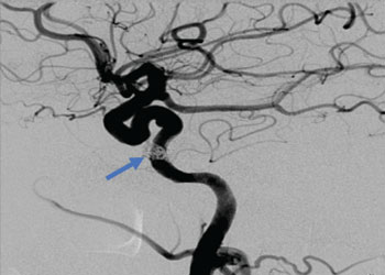
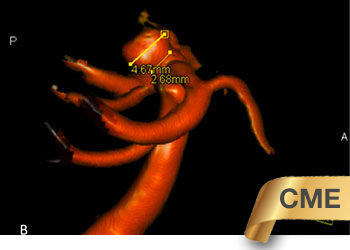
Carotid Cavernous Fistula (CCF) Study
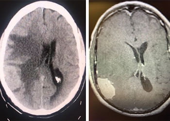
October 27, 2021MRI with gadolinium confirmed the likely diagnosis of convexity meningioma, a benign neoplasm of the meninges. Given the location of the tumor, angiography and embolization were deemed unnecessary prior to resection. Stereotactic c-guided craniotomy was performed by Dr. Jonathan Brisman and the tumor was carefully excised from the surrounding brain. A gross total resection was achieved. He was discharged from the hospital to his home, neurologically intact with a slow improvement of his gait. Postoperative MRI showed no evidence of residual tumor.

Pathologic analysis revealed a meningothelial tumor consistent with a meningioma with atypical features (immunocytochemical positive staining for vimentin, EMA, and Ki67 at 23%), and the lesion was graded an Atypical Meningioma, WHO grade 2. Neuro-oncology consultation was obtained and a decision was made to monitor conservatively, with radiation therapy indicated for tumor recurrence only. The patient has since returned to normal neurologic function with no evidence of tumor recurrence
CATEGORY: BRAIN // ATYPICAL CONVEXITY MENINGIOMA
Atypical Convexity Meningioma
The patient is an otherwise healthy 80-year-old gentleman who presented with complaints of right frontal headaches and difficulty walking that had progressed over about three weeks. He was neurologically nonfocal. CT scan without contrast showed a large right parietal mass with midline shift and cerebral edema.