- PATIENT FORMS | REQUEST A CONSULTATION | CONTACT US
- 1-844-NSPC-DOC
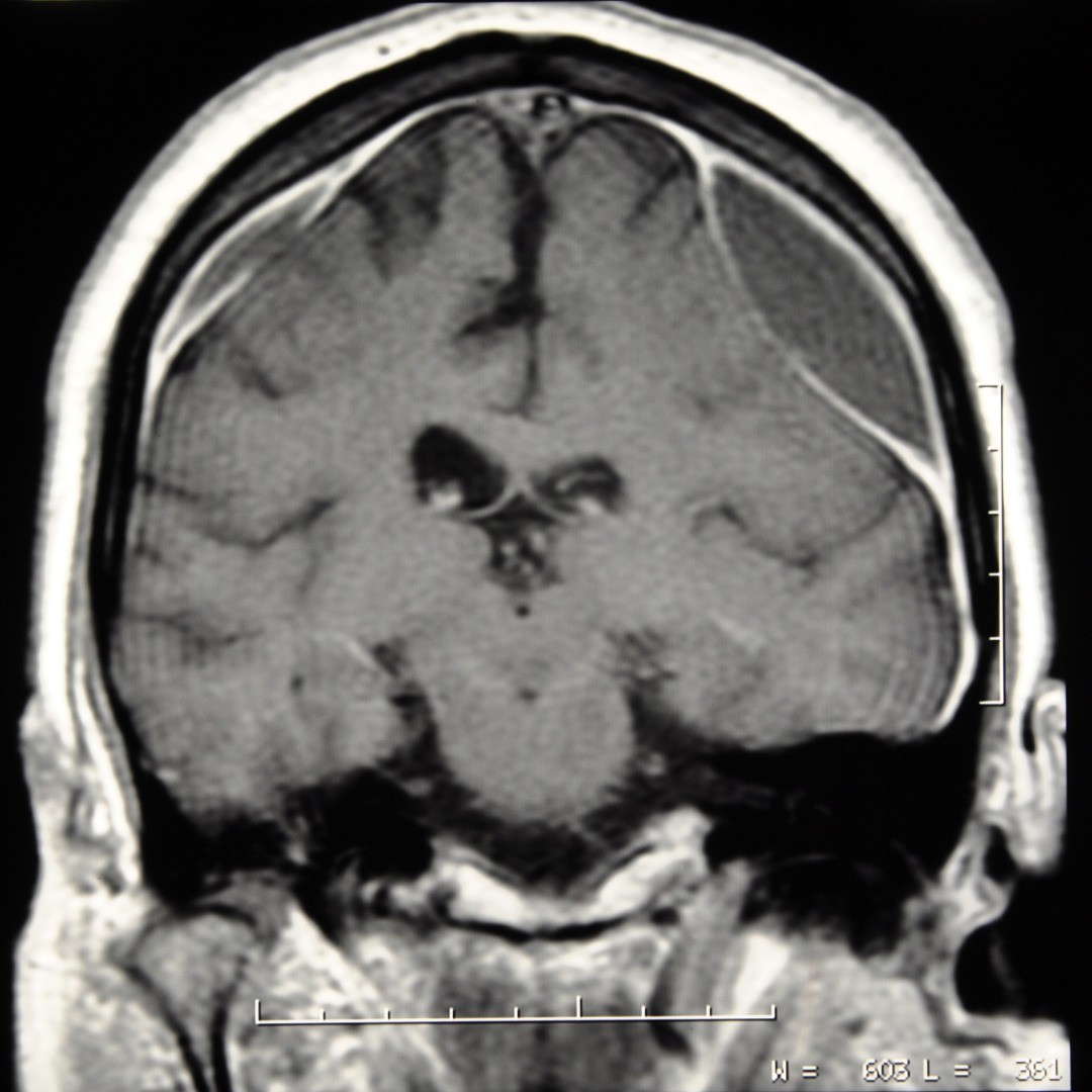
Craniotomy for Subdural Hematoma
A craniotomy for subdural hematoma allows a neurosurgeon access through the skull via a small opening for the extraction of a blood clot on the exterior of the brain. These blood clots (hematomas) are underneath (sub) the dura mater (dural) or outer covering of the brain and occur when the blood vessels that traverse the space are ruptured.
Subdural Hematoma Symptoms
The symptoms for a subdural hematoma resulting from a head injury can be immediate, known as an acute subdural hematoma, or can develop over time after an injury—sometimes even several weeks, called a chronic subdural hematoma. Symptoms can include:
- a headache which may be continual or ebb and flow
- differently sized pupils
- blurred vision
- nausea and/or vomiting
- lethargy
- weakness on one side of the body
- discernible head wound
- slurred speech
- confusion
- loss of consciousness
- seizures
A cerebral hematoma requires emergency medical attention to prevent neurological damage or death. At NSPC (NSPC Brain & Spine Surgery (NSPC)), we have neurosurgeons on call to take doctor referrals for emergency brain surgery in the New York area.
What Is the Procedure for a Craniotomy for Subdural Hematoma?
This open brain surgery requires general anesthesia of the patient. Some of the scalp is shaved and special pins are used to guard the head against movement.
The neurosurgeon carefully makes an incision in the skin above the brain hemorrhage. Small burr holes are drilled to allow the insertion of microsurgical saws to create a temporary opening in the bone. The bone flap is removed, creating a small window, and set aside to be attached later.
Through this small window, the dura is carefully incised to provide an entrance to the hematoma. Burr hole surgery may also be used to divert excess fluid away from the brain, which can decrease cerebral pressure and avert possible brain damage.
A special surgical device gently suctions the excess blood, and if necessary, any additional bleeding is cauterized.
Once the subdural hemorrhage has been cleared, the dura is sutured and the small piece of skull reattached with surgical pins and screws. If necessary, the reattachment of the skull piece may be postponed and a drain may be temporarily located at the incision to prohibit accumulations of fluids. The scalp skin is then sutured closed.
Hospitalization after this cerebral hematoma treatment may be as short as a few days, but can sometimes be longer depending on where the brain hematoma was and its size.
Physicians
Connect With Our 7 Convenient Locations
across Long Island, NY
Our expert physicians, surgeons and doctors are ready to serve you at our 7 convenient locations across Long Island, NY. Connect today to learn how our award winning, world class experts can help.
4250 Hempstead Turnpike Suite 4,
Bethpage, NY 11714
(516) 605-2720
COMMACK
353 Veterans Memorial Hwy,
Commack, NY 11725
(631) 864-3900
One Hollow Lane, Suite 212
Lake Success, NY 11042
(516) 442-2250
MANHATTAN
215 E. 77th Street Ground Floor
New York, NY 10075
(646) 809-4719
EAST SETAUKET
226 North Belle Mead Road, Suite C
East Setauket, NY 11733
(631) 828-3001
100 Merrick Road, Suite 128W
Rockville Centre, NY 11570
(516) 255-9031
WEST ISLIP
500 Montauk Hwy
West Islip, NY 11795
(631) 983-8400
World
Class
Expertise
For over 50 years & 350,000 patients NSPC has been a trusted global medical leader.
Contact us today for an appointment or consultation.


