- PATIENT FORMS | REQUEST A CONSULTATION | CONTACT US
- 1-844-NSPC-DOC
Posterior Fossa Brain AVM – 20-Year Follow-Up

Revision Surgery Relieves Woman’s Chronic Low Back Pain Case Study
July 21, 2022
Patient with Cervical Spine Trauma Suffers Multiple Concurrent Pathologies
March 26, 2022A 40-year-old woman who was 5 months pregnant, presented to the emergency room with a sudden onset of a bad headache. CT and MRI of the head showed a small bleed from a posterior fossa arteriovenous malformation (AVM) in the superior portion of the cerebellar vermis. After a few days, she felt better and was discharged. Several months later she completed her pregnancy with a healthy delivery.
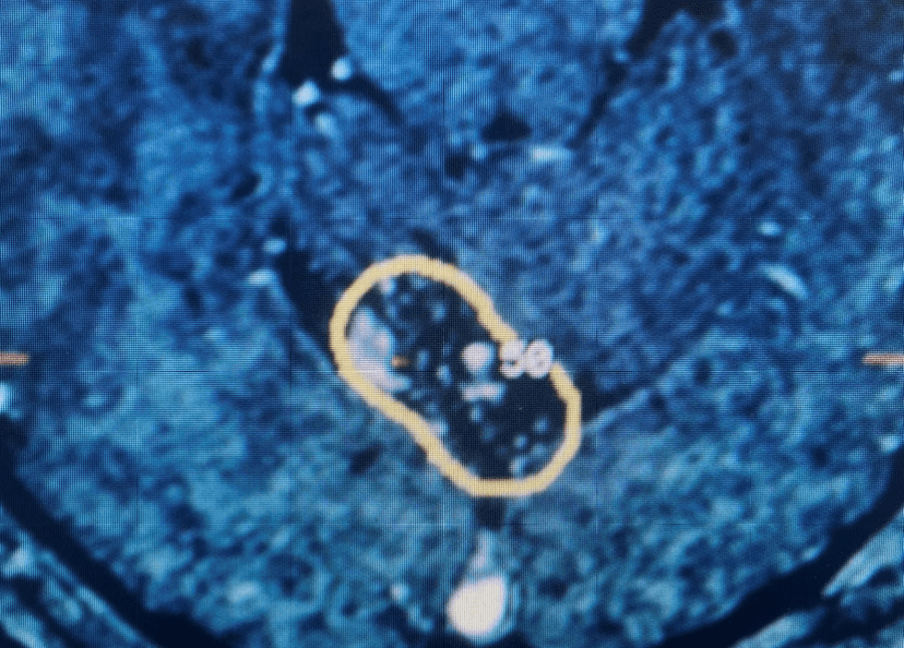
Subsequent to this, she underwent Gamma Knife treatment of the cerebellar AVM (figure 1) with Dr. Michael Brisman. She had no subsequent problems. Subsequent imaging showed that the AVM was starting to thrombose. She was then lost to follow up for many years.
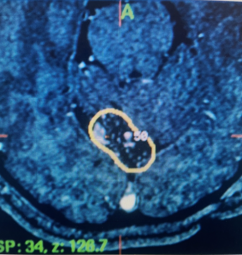
Figure 1. Axial MRI with contrast at the time of gamma knife treatment showing the tightly conformal 50% Isodose line contoured around the AVM of the posterior superior cerebellar vermis.
She returned for a follow up visit 19 years after the Gamma Knife treatment just to check up on things. She felt well and was neurologically intact. MRI and CTA of the brain were performed, which demonstrated complete obliteration of the AVM.
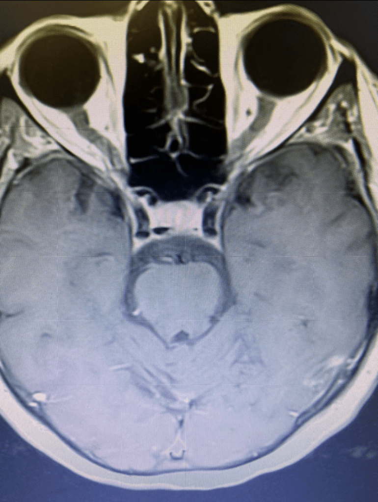
Figure 2A. Axial MRI with contrast.
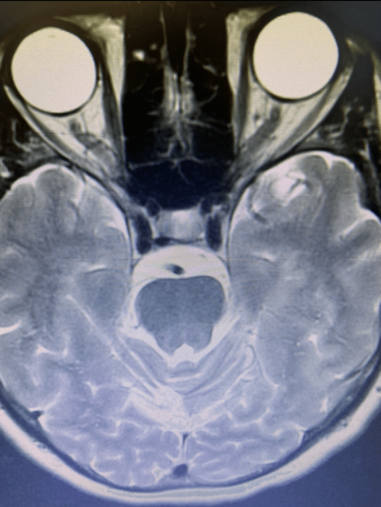
Figure 2B. Axial T2 MRI.
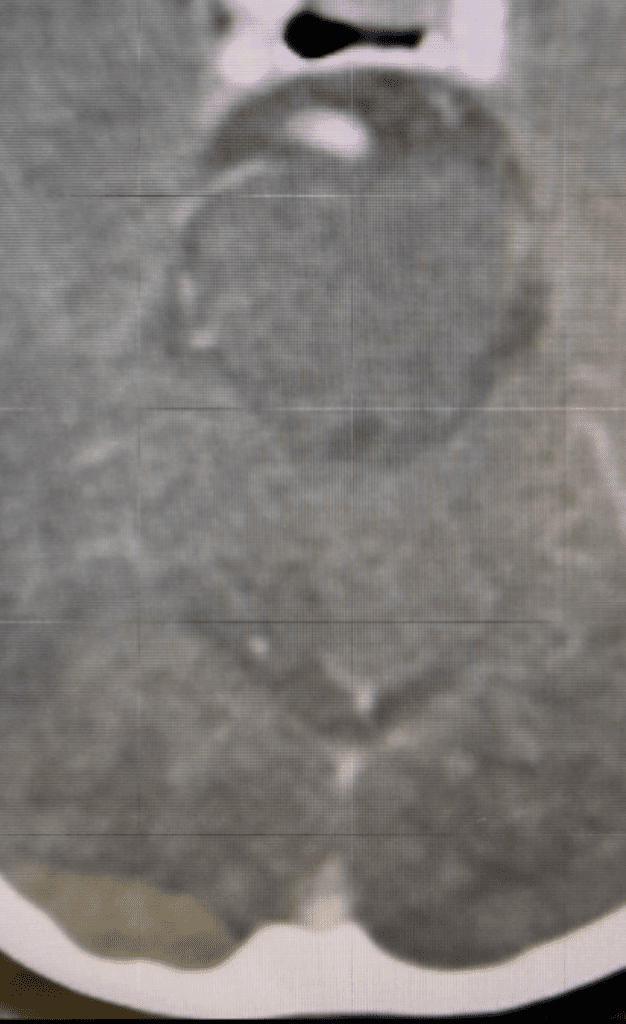
Figure 2C. Axial CTA image.
Discussion:
Brain AVM’s are abnormal tangles of blood vessels that are usually congenital. They usually present with seizures or bleeding within the brain. The annual bleed rate is about 3%, and about half of these will lead to death or major disability.
Very small Brain AVM’s can be obliterated with embolization. Very large AVM’s should be managed without any procedure. Surgery can be considered in small superficial AVM’s that have bled, that occur in young, healthy patients, are in non-eloquent brain, and can be well embolized before the surgery. Most Brain AVM’s are best treated with a one day, out-patient treatment with Gamma Knife.
The super-focused radiation of Gamma Knife usually causes the AVM to thrombose over 2-4 years. If there is any residual, a repeat treatment can be performed years later to treat the remaining portion. Some larger AVM’s can be treated in stages, treating part of the AVM with Gamma Knife in one sitting, and then the remaining portion at a later date.
This patient was a young healthy patient with a moderate sized AVM in the posterior fossa that had bled. As such, treatment with Gamma Knife was appropriate. It was also appropriate to wait a few months for her to complete her pregnancy.
We usually perform Gamma Knife with concurrent MRI and Angiographic imaging. Gamma Knife allows for a very conformal dose of radiation right to the center (“nidus”) of the AVM.
This patient had an uncomplicated treatment of her brain AVM, that successfully obliterated the AVM, and showed excellent long term follow up, at 19 years. The AVM disappeared and she never had any further symptoms.
CATEGORY: BRAIN // POSTERIOR FOSSA BRAIN AVM – 20-YEAR FOLLOW-UP
Posterior Fossa Brain AVM – 20-Year Follow-Up