- PATIENT FORMS | REQUEST A CONSULTATION | CONTACT US
- 1-844-NSPC-DOC
50 year old man with new onset aphasia and a left frontal mass / Brain Abscess
Laminectomy and Fusion Solves Decades of Back Pain
October 27, 2021
Hemangioblastoma
October 27, 2021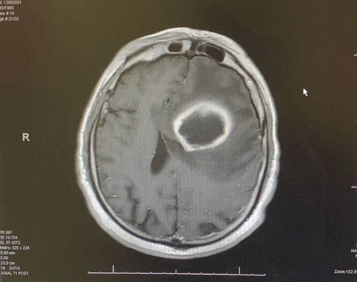
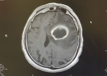
Image 1: Pre-treatment axial post contrast MRI.
Because the lesion was sizeable, and causing much mass effect, and the diagnosis was unclear, and the patient was young with no other existing disease, it was decided to emergently bring the patient to the operating room for surgical removal of the mass. Surgery was performed by Dr. Michael Brisman. Stereotactic Brainlab navigation was utilized. A small left frontal craniotomy was performed. After making a small cortical incision, the Vycor transparent tubular retractor was inserted into the lesion. Thick yellow liquid was encountered under pressure consistent with frank purulence. (Image 2 shows intra-operative image of Vycor tubular retractor in deep left frontal mass with thick purulent material coming out under pressure.) Cultures were sent. The pus was fully washed out with gentle antibiotic irrigation.
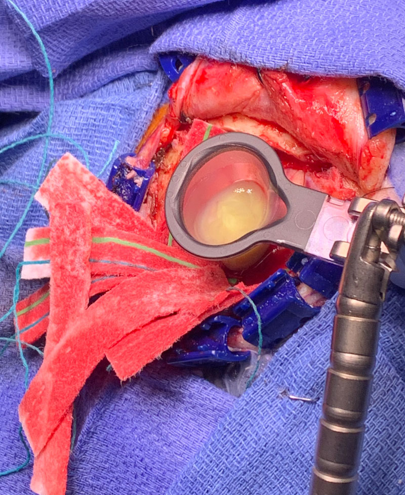
Image 2: Intra-operative image of Vycor tubular retractor in deep left frontal mass with thick purulent material coming out under pressure.
Postoperatively, the patient was started on broad spectrum IV antibiotics, under the supervision of an Infectious Disease consult. Clinically, the patient gradually recovered. Within weeks, the patient had fully recovered and was neurologically intact. Gram stains were suspicious for the presence of bacteria (encapsulated cocci). Cultures never grew out any organisms. The patient was treated with several weeks of broad spectrum IV antibiotics. The patient made a full recovery. (Image 3, axial post contrast MRI from 3 months post-op shows resolution of brain abscess and surrounding edema. )
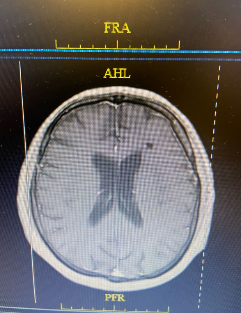
Image 3: Axial post contrast MRI from 3 months post-op shows resolution of brain abscess and surrounding edema.
Brain abscess can result from direct spread, from hematogenous spread, and from penetrating injuries. In this case, the abscess was presumably from hematogenous spread. The source in this patient was never identified. Different organisms can cause a brain abscess, and abscesses can be of variable size. They can also be single or multiple. General treatment is with various antimicrobial agents. Indication for surgical intervention of a brain abscess would be (1) to confirm the diagnosis, including identifying the organism and (2) to decompress a large lesion. Urgent surgical evacuation was indicated in this case to establish the diagnosis and to decompress a large very symptomatic lesion. Surgery in this case was life saving and curative.
CATEGORY: BRAIN // BRAIN ABSCESS
50 year old man with new onset aphasia and a left frontal mass / Brain Abscess
This is a 50 year old man who last year had undergone gastrectomy for gastric cancer. He presented now with new onset of lethargy and aphasia, with both a receptive and expressive component . He was also experiencing intermittent bradycardia. Brain imaging showed a 3 cm ring enhancing , fluid filled mass in the left frontal region with significant surrounding edema, mass effect, and midline shift. (Image 1 shows axial post contrast MRI. ) Recent imaging of the body had shown no evidence of active metastatic disease.