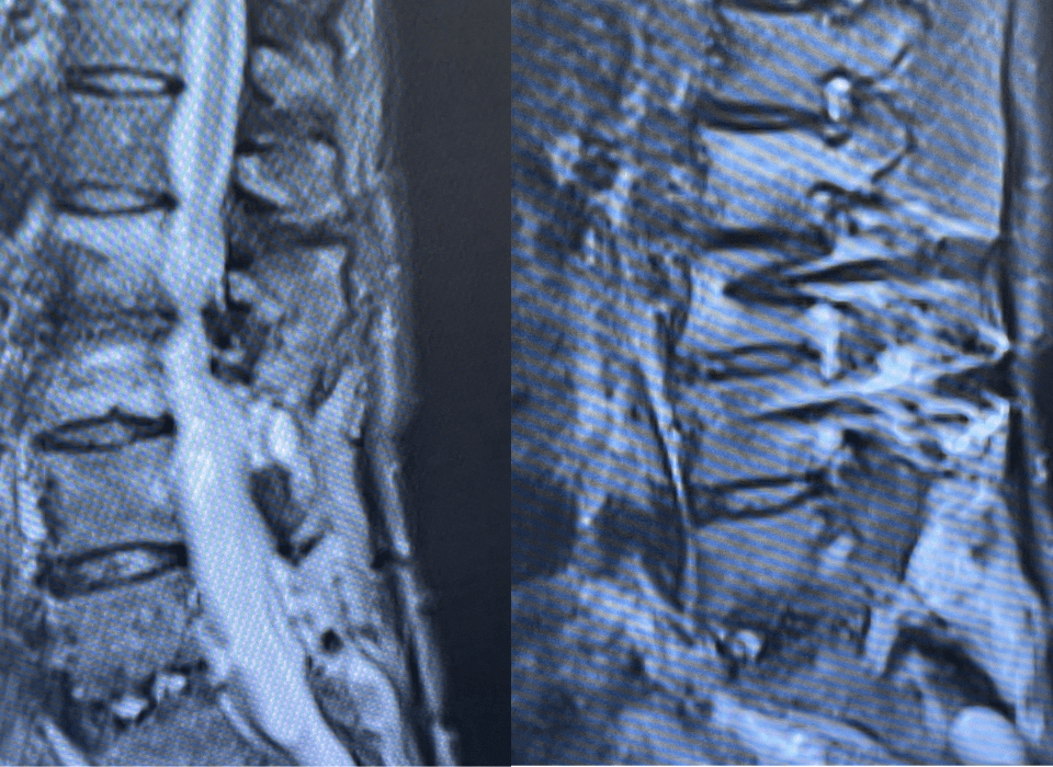
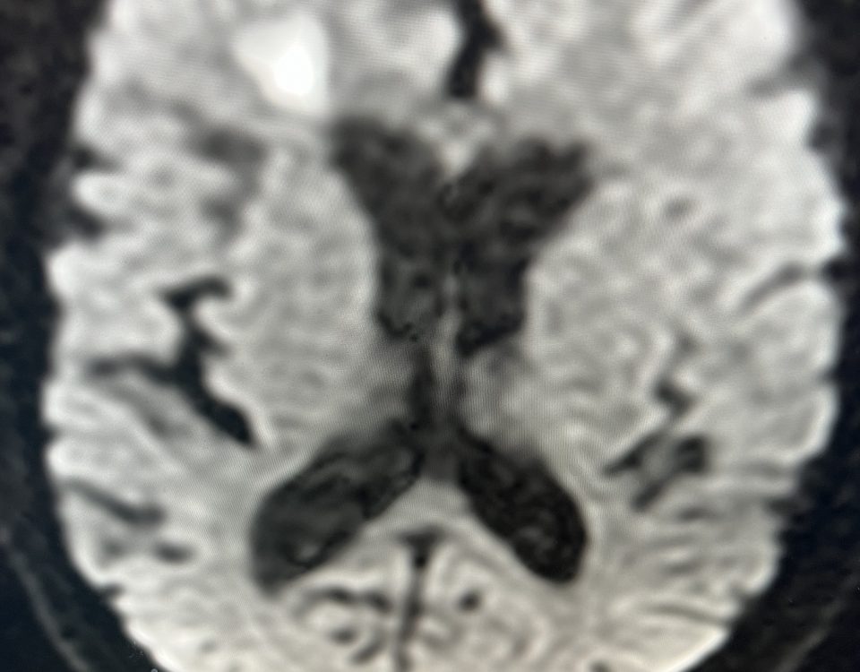
First, the diagnosis of a complex migraine was made in this young mother. A brain MRI on presentation revealed an evolving right parietal infarct (Fig 1). Furthermore, the brain MRA showed bilateral distal internal carotid and right vertebral artery narrowing suggestive of arterial dissections. The distribution of the stroke was highly characteristic of deep white matter and cortical watershed areas of the ACA/MCA/PCA territories (“Triple Watershed”).
Figure 1. Brain MRA disruption of vascular filling in bilateral internal carotid and vertebral arteries suggesting multi-vessel dissections (A). Right parietal watershed area infarct (yellow arrow) – subacute on diffusion weighted imaging (B) and FLAIR (C).
She was admitted to the hospital and underwent digital subtraction catheter angiography (Fig 2) which confirmed the extent and severity of the dissections and long-segment stenoses in all four pre-cerebral arteries. It also revealed a left vertebral artery flap. A work-up for vasculitis was negative.
Figure 2. Baseline DSA angiogram showing dissection in all four pre-cerebral arteries (blue circle): right vertebral artery (A); right internal carotid artery (B); left internal carotid artery (C); left vertebral artery (D). Extracranial to intracranial collateralization (E, F) and posterior-to-anterior collateralization (D, G) with filling of the internal carotid artery circulation via the posterior communicating artery (D) suggesting shifting perfusion.
She was placed on Aspirin and Coumadin and followed with brain MRA at 3 months that demonstrated continued improvement and stabilization of the arterial dissections under medical therapy (Fig 3).
Figure 3. Neck and brain MRA (A, B) at 3 months. Stable bilateral internal carotid artery and vertebral artery dissections with significantly improved vascular filling of same vessels. Baseline MRA as comparison.
This was followed by another brain MRA at 6 months and angiography at 8 months which confirmed ongoing stability, improved flow, and healing dissections (Figure 4).
Figure 4. Right (A) and left (B) internal carotid artery angiogram and right (C) and left (D) vertebral artery angiogram at 8 months with improved flow. Stable arterial dissections and caliber changes (red arrows). Compared to baseline, reduced posterior-to-anterior collateralization.
She remained asymptomatic without recurrent ischemic events — for one year …
Discussion:
Multiple simultaneous cervical artery dissections are rare and have been typically associated with trauma[1] or connective tissue disorders[2]. In most cases, however, no underlying cause can be identified, as in our young woman.
In patients under 45 years of age, cervical-cranial artery dissections cause approximately 1/4 of strokes. Medical treatment is highly effective in routine cases of such extra-cranial dissections. Antiplatelet medications have become the mainstay of treatment, replacing oral anticoagulation.[3] It is often practiced to use both for a short period of time before bridging to antiplatelet therapy long-term. In cases where continued flow-failure remains an issue, as in this patient, continuation with combined oral treatment for longer periods may be beneficial.
Acute angioplasty and stenting are rarely recommended for cervical dissections, unless as part of treating an intracranial large vessel occlusion (LVO) related to an acute stroke syndrome.
MRI and MRA offer an excellent non-invasive way to follow these patients with a sensitivity greater than 90% in most studies. There are, however, indications for complementary contrast enhanced imaging (CTA, DSA), e.g., to better clarify severity of stenosis and to monitor flow. MR imaging is less sensitive in vertebral artery dissections, especially in segments with tortuosity.[4]
Recurrent stroke risks for isolated cervical Carotid or Vertebral Artery dissections on antiplatelet or anticoagulation therapy have been observed to be very low at 1 year, with multicenter randomized prospective studies reporting an incidence of approximately 2.5% on medical therapy (CADISS Trial).[5]
Our patient’s initial clinical course progressed as we hoped and expected … for one year, however, patients do not always follow scripts, and her story is no exception…
References:
Her story started almost five years ago. A woman in her 30s experienced neck pain and headache that had been bothering her for a month. Without any preceding trauma, she was attributing it to her hunching over her computer. One morning, she became nearly mute, unable to find words with weak left arm and hand.
To learn more about the author, click his name or photo: