Educational Goals:
Learners will be able to recognize the symptoms that may suggest a Left MCA occlusion stroke syndrome, order appropriate tests to confirm the diagnosis, and appropriately refer these patients to a subspecialist for management and treatment.
“90’s-year-old female with past medical history of hypertension, hyperlipidemia, atrial fibrillation on daily aspirin brought in by EMS as family noticed patient stopped speaking and could not hold her coffee cup at approximately 9:50 AM today. Has been nonverbal since. No trauma or falls. Last known normal prior to that as she walked to the kitchen table.”
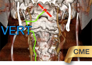
Upon evaluation in the Emergency Department, she was found to have an NIH Stroke Scale score of at least 15, with global aphasia, right hemiparesis, neglect, sensory deficit, and fixed eye deviation compatible with Left MCA Syndrome. Emergent non-contrast CT reveals remote small left parietal infarct, while CT Angiography confirms a left ICA/M1 occlusion (Fig. 1A) with corresponding CT Brain Perfusion of at-risk left MCA territories and mis-matched regional defects representing spectrums of ischemia (Fig. 1B).
Patient arrived < 1 hour from event and received IV tPA per hospital stroke protocols. Despite her early presentation, a mixture of matched and mis-matched defects suggesting varied levels of ischemic progression were observed on perfusion imaging. She was brought emergently for Mechanical Thrombectomy.
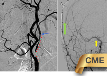
A Reverse Curve Vitek 125 cm 5 French Catheter was used to guide the 6 French Shuttle across tortuous Aortic Arch, Thoracic, and Cervical Carotid vasculature (Fig. 2A). With the 6F Guide Sheath as a stable platform, the left ICA/MCA occlusion was crossed with a microwire (Fig. 2B) and the .064 Penumbra Reperfusion catheter was position for First Pass Aspiration Thrombectomy (Fig. 2C). Aspiration thrombectomy using the ADAPT technique was performed with negative aspiration of the thrombus for approximately 5 minutes after which time the reperfusion catheter was slowly withdrawn with negative aspiration of the left ICA guiding sheath contemporaneously.
Post thrombectomy angiography demonstrates reperfusion of 60-70% of the left MCA territory with distal M2 embolus in Inferior Parietal-Occipital Branches (Fig. 3).
A 4 Max reperfusion catheter and 4 Max Separator were used to perform additional aspiration thrombectomy resulting in complete reperfusion (Fig. 4).
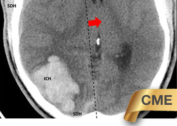
Within 24 hours, she had near complete recovery of language and strength (NIHSS 2-3) with 48 hour CT demonstrating mild edema within the external capsule and subinsular cortex (Figure 5).
She celebrated another milestone birthday in the hospital and was soon home with family!
Discussion:
Although mechanical thrombectomy has become the standard of practice for Large Vessel Occlusions in Acute Stroke, many challenges remain. Among them, selecting patients that are most likely to benefit and expanding care to these populations. With the rapid development of efficient Stroke Triage and Imaging Paradigms, we have been successful in better identifying patients suffering from a disabling or life-threatening stroke, as well as beginning to explore the prognostic value of clinical and imaging phenotypes and the effects of treatment.
Advanced age, by design, has often been considered an exclusion in most landmark clinical trials (NINDS, MR CLEAN, others.)1,2,3 As our experience grows, we are learning that many effective treatments can be considered even in the elderly (>89)4. People who survive into their 90’s often have years of functional life-expectancy (LE) remaining (90-99yo = 4.05-2.24 yLE),5 having self-selected by their genetic, epigenetic, and constitutional survivor bias. At age 90, a person has > 4 years of LE, while at age 99, LE is > 2 years. Although most clinical trials, by design, exclude patients at the extremes of age (<18, >89) and LE (< 6 months), pre-morbid functional status, comorbidities, clinical, and imaging presentations should be considered and may extend significant benefits to these patients. This patient’s constitution and favorable perfusion profile with successful reperfusion returned her to a pre-stroke Baseline (mRS-2) at Discharge with another >2 years of LE projected.
Stroke Imaging Systems have rapidly evolved to allow for timely diagnosis and physiologic assessments of brain perfusion and potential opportunity. CTA imaging rapidly identifies the sites of occlusion, collaterals, and the navigation pathway. CT and MR perfusion imaging provide a physiologic snapshot of the dynamic ischemic cascade6. Many studies have begun to develop favorable perfusion phenotypes, as well as those of “futility”. Our experiences have confirmed that patients often experience significantly smaller strokes after successful reperfusion therapy and improved functional recovery. Nevertheless, despite early and complete revascularization in many MCA occlusions, we often observe injury within the basal ganglia and sub insular cortex. This raises many interesting questions on the ischemic tolerance of different tissues and territories based on location of occlusions, their timing, collaterals, and metabolic activity perhaps related to shifting of the ischemic watershed to these regions. This territory seems particularly vulnerable.
A combination of classic techniques and next-generation medical devices continues to result in improved rates of revascularization. Access to the occlusion can now be approached from femoral, radial, and direct carotid approaches – mapped, planned, or improvised. Mechanical thrombectomy techniques now include a wide-breadth of strategies (Aspiration, Stent-Retrievers, Balloon Guides) which have resulted in high rates of reperfusion (60-90%). As the application and experience of Endovascular Revascularization Therapy (ERT) has grown within Large Vessel Occlusions LVOs (ICA, M1, M2, Basilar), many Neurovascular Teams have carefully selected more distal occlusions to consider for revascularization. Although we achieved reperfusion in the anterior 2/3’s of her left MCA on First Pass, we chose to pursue aggressively her parietal-occipital M3 embolus in an effort to preserve language and its devastating effects in the elderly7. Often decisions to proceed with additional intervention have to be carefully weighed with the risks and the benefit to each patient, with careful consideration of the patient, family, and community. The volume of the stroke may be significantly reduced with near-complete revascularization of LVOs, however, persistent distal occlusions of M2-M3 branches in highly eloquent locations (Left Hemispheric Language or Rolandic Motor) may result in severe functional disability and may be considered as targets for ERT by experienced Neurointerventionalists<sup8.
References:
Disclosures:
The planners and faculty participants do not have any financial arrangements or affiliations with any commercial entities whose products, research or services may be discussed in these materials.
CME Accreditation:
This activity has been planned and implemented in accordance with the accreditation requirements and Policies of the Medical Society of the State of New York (MSSNY) through the joint providership of the Academy of Medicine of Queens County and NSPC Brain and Spine Surgery. The Academy of Medicine of Queens County is accredited by The Medical Society of the State of New York (MSSNY) to provide continuing medical education for physicians. The Academy of Medicine of Queens County designates this Enduring Materials for a maximum of 1 AMA PRA Category 1 Credits™ as specified in this activity. Physicians should claim only the credit commensurate with the extent of their participation in the activity.