Dural Arterial Venous Malformations

Grade II Oligodendroglioma
October 27, 2021
Unruptured Brain Aneurysms of the Middle Cerebral Artery
October 27, 2021

Deep venous drainage is observed into the Galenic and Straight Sinus of the posterior fossa. Successful trans-arterial embolization with liquid embolic (N-butyl cyanoacrylate, NBCA) of the bilateral external and left vertebral posterior meningeal artery feeders was successful at reducing >90% of these high-pressure indirect shunts (Figure 1D), with small residual shunts from the distal occipital arteries that were subsequently targeted for cure with Gamma Knife Radiosurgery.

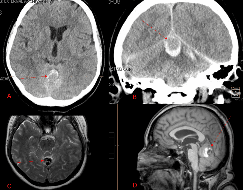
Figure 1. [A] Ruptured aneurysm of the Deep Venous Posterior Fossa drainage and AV shunting [B] Posterior Meningeal Artery Fistula [C] Right Middle Meningeal Artery Fistula [D] Post embolization Left Vertebral Angiogram demonstrates resolution of the high flow and pressure AV Shunting following successful embolization with liquid embolic (NBCA).

Key Learning Points:

  1. Indirect Dural AV Fistula Vascular Malformations represent high risk lesions that result from acquired and/or congenital indirect fistula and A-V shunts that can arise within many of the deep venous structures of the brain (Superficial and Deep Venous Drainage systems).
  2. Many patients may present with initially mild symptoms of pulsatile tinnitus or headaches but may progress over time to more severe high-risk symptomatology including hemorrhagic stroke, permanent neurologic deficits, and even mortality.
  3. Early evaluation by an expert multi-disciplinary team is critical to diagnosis, management, and successful treatment and recovery with a variety of medical, endovascular, and surgical approaches.”

REFERENCE:
Management of tentorial dural arteriovenous malformations: transarterial embolization combined with stereotactic radiation or surgery. Lewis A, Tomsick T, Tew J. Journal of Neurosurgery. Volume 81: Issue 6 (Dec 1994). Issue 6 (Dec 1994). Here is the link https://pubmed.ncbi.nlm.nih.gov/7965115

CATEGORY: ENDOVASCULAR // DURAL ARTERIAL VENOUS MALFORMATIONS

Dural Arterial Venous Malformations

A 53-year-old man presented with acute onset of severe headache, and possible witnessed seizure. Imaging confirmed intraventricular hemorrhage primarily focused within the fourth ventricle with diffuse subarachnoid hemorrhage within the posterior fossa and along the tentorium (Figure 1A). No severe neurologic deficits were observed initially. His cerebral angiogram revealed a high flow dural arterial-venous fistulous malformation of the tentorium supplied by the bilateral external carotid and left vertebral arteries into a large venous varix within the fourth ventricle at the site of the hemorrhage (Figure 1B, C).

AUTHORS:
To learn more about the author, click his name or photo:
Share

Authors

To learn more about the author, click his name or photo: