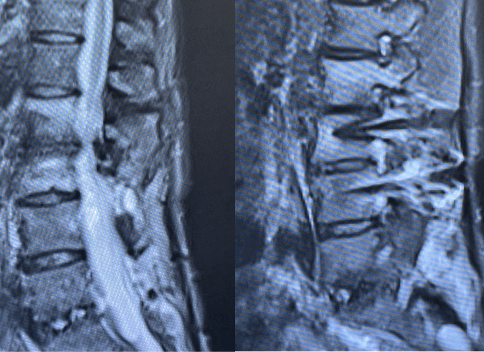
Figure 1: Spinal MRI showing the collapse of T9 vertebral body with epidural tumor (*) pressing on the spinal cord (M) and causing significant cord edema.
This 73-year-old woman presented with weakness of her right leg and perineal numbness. A cancer survivor, she was known to have renal cancer. A hypervascular tumor to T9 with mass effect and spinal cord edema was found (Figure 1). With the presumed diagnosis of metastases from renal cell CA (Figure 2) to the T9 epidural region, the hypervascular tumor was expected to be located where the anterior spinal artery is often seen. In this difficult situation, embolization and surgery were offered to the patient with the modest goal of keeping her ambulatory as long as possible. The patient was scheduled for pre-operative embolization and surgery.
Figure 2: Renal Cell Carcinoma are extremely vascular tumors with intricate and arborizing vasculature.
The main goal of the embolization was to devascularize the tumor to make the surgery safer by reducing intra-operative blood loss. Additionally, an initial angiogram could help assess whether the anterior spinal artery was at risk by embolization or surgery.
Super-selective angiography provided sufficient information to exclude the risk of embolizing or damaging the anterior spinal artery (Figure 3). Additionally, an anesthetic agent opacified with contrast was subsequently injected into vessels with intra-operative monitoring, Motor- and Sensory-Evoked studies, that confirmed there were no angiographically-occult arterial feeders.
Pre-operative embolization was performed with osmotic agents under general anesthesia and with intra-operative monitoring (Figure 4). This was followed by proximal occlusion with PVA and coil occlusion of the intercostal arteries adjacent to the take-off from the aorta which led to total obliteration of the tumor (Figure 5). The patient was clinically stable after the embolization. The surgeon reported a completely avascular tumor. The patient improved neurologically.
Figure 3: AP view of T9 intercostal angiogram showing massively vascular tumor involving the pedicle and vertebral body.
Figure 4: AP magnified view of T9 inter-costal angiogram post-embolization with osmotic agents alone and prior to finishing with a small amount of PVA and coils within the proximal intercostal artery. Near-complete obliteration of the tumor.
Figure 5: Post-operative T9 vertebra with widely open adjacent spinal canal (yellow circle).
Treatment Considerations:
Endovascular Osmotic Embolization With routine embolizing agents, such as PVA or Embospheres™, as well as liquid agents, such as Onyx™ and Truefill™, immediate postembolization cytotoxic and vasogenic edema or hemorrhage occur frequently and can cause spinal cord damage. Additionally, inadvertent embolization of adjacent feeding collaterals to the spinal cord with particles or solidifying liquid agents is problematic.
Additionally, a complete devascularization with routine embolization agents is often difficult in hypervascular tumors due to incomplete penetration. Chemotherapeutic and radioactive embolic agents partly address the heterogeneous penetration problem of the particles or liquid emboli but would not be useful to devascularize the tumor acutely.
Osmotic embolization was pioneered to overcome the technical limitations of other embolization agents. It can be used for palliative, adjunct, and definitive treatment of tumors. Its major advantage is deep penetration into the vascular territories and tissue death by terminal dehydration with subsequent shrinkage. It is painful and monitored anesthesia care or general anesthesia is required.
REFERENCES:
Feng L Kienitz BA, Matsumoto C, Bruce J, Sisti M, Duong H, Pile-Spellman J. Feasibility of using hyperosmolar mannitol as a liquid tumor embolization agent. AJNR 2005; 26: 1405-12.
To learn more about the author, click his name or photo: