Figure 1. Head CTA showing an aneurysm in the pericallosal branch of the left anterior cerebral artery (circle).
A woman in her sixties was admitted with mental status change. Brain imaging (CT/CTA) found a hyperdense mass in the pericallosal branch of the left anterior cerebral artery (Figure 1). The patient was diagnosed with atypical bacterial pneumonia and treated with antibiotics per antibiogram. Ten days later and in stable condition, she was discharged with extended antibiotic treatment. Having been seen early in the process by the Neurovascular Team of NSPC, the patient was scheduled to follow-up with the Neurovascular Team regarding the pericallosal mass that was suggestive of a mycotic (infectious or septic) aneurysm.
Control brain MRA confirmed an irregularly shaped left pericallosal branch aneurysm (Figure 2).
Figure 2. Brain MRA 3D-time-of-flight showing a 7mm x 5mm aneurysm in the pericallosal branch of the left anterior cerebral artery (asterisk).
Based on the arterial site of the aneurysm and underlying infection (pneumonia), it was believed to be a mycotic aneurysm. Due to the size and persistence of the mycotic aneurysm, eradiation treatment was considered. Following, a conventional angiogram and endovascular embolization of the aneurysm was planned. Baseline cerebral angiogram found an irregularly shaped pericallosal saccular aneurysm that had increased in size over the past weeks (Figure 3).
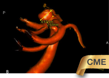
Figure 3. Cerebral angiogram, lateral view (top) shows contrast-enhancing mass on the left pericallosal branch suggestive of a mycotic aneurysm (asterisk).
3D-reconstruction, lateral and craniocaudal views (bottom), shows a saccular aneurysm with an irregular shape (lobulated) of about 6mm x 9mm x 3mm in dimensions.
Although the aneurysm was asymptomatic, treatment was indicated due to the dynamic anatomy and infectious nature of the aneurysm. Embolization was performed with coils and resulted in near complete obliteration of the aneurysm (Figure 4). The patient had an uneventful course.
Figure 4. Post-coiling cerebral angiogram with near-complete embolization of the pericallosal branch aneurysm (circle, left). Coils can be seen within the aneurysm sack (circle, right).
Treatment Considerations
Mycotic or infectious aneurysms of the brain represent a minority (<5%) and may occur with systemic or local spread of an infection to a cerebral artery [1,2]. Most frequently, the cause is a bacterial infection (other causes include fungi and viruses) that can involve local and distant structures, such as the meninges, periorbital structures, sinus veins, endocardium, and lungs [1]. Infection and inflammation of the arterial wall lead to degenerative remodeling processes, thereby weakening the vessel wall integrity, ultimately allowing an aneurysm to form.
Typically, mycotic aneurysms are located in the periphery unlike true aneurysms for which more proximal cerebrovascular junctions are predilection sites. However, mycotic aneurysms may be located on proximal cerebral vessels. Due to the infected, inflamed and weakened vessel wall, mycotic aneurysms are prone to rupture with likely detrimental outcome for most of the affected.
The primary therapy is treatment of the underlying infectious cause with extended antibiotic treatment. Mycotic aneurysms should be closely watched by professionals with neurovascular expertise for regular assessment of the risk of rupture and treated when size remains unchanged or increases [3,4,5].
The lack of randomized clinical trials has prevented a definition of a standard in practice. The identification and treatment of the infectious cause and careful observation of the mycotic aneurysm with timely effective intervention to eradicate the lesion appear prudent practice.
References
Disclosures:
The planners and faculty participants do not have any financial arrangements or affiliations with any commercial entities whose products, research or services may be discussed in these materials.
CME Accreditation:
This activity has been planned and implemented in accordance with the accreditation requirements and Policies of the Medical Society of the State of New York (MSSNY) through the joint providership of the Academy of Medicine of Queens County and NSPC Brain and Spine Surgery. The Academy of Medicine of Queens County is accredited by The Medical Society of the State of New York (MSSNY) to provide continuing medical education for physicians. The Academy of Medicine of Queens County designates this Enduring Materials for a maximum of 1 AMA PRA Category 1 Credits™ as specified in this activity. Physicians should claim only the credit commensurate with the extent of their participation in the activity.
To learn more about the author, click his name or photo: