- PATIENT FORMS | REQUEST A CONSULTATION | CONTACT US
- 1-844-NSPC-DOC
Treatment of Spondylolisthesis and Disc Herniation
Unusual Indication for Carotid Stenting Over Endarterectomy
May 2, 2022
Surgical View of a Microvascular Decompression (MVD) for Trigeminal Neuralgia
May 27, 2022This patient is a 63-year-old man who has suffered with lower back pain and right leg pain for several years. His pain tends to increase with standing and ambulation. When his pain initially began, he got relief with over-the-counter medications (i.e. Advil, Tylenol). He then began to receive epidural steroid injections, which lasted for a while but the pain soon returned.
His MRI lumbar spine showed a herniated disc at L2/L3 and a grade 1 spondylolisthesis at L4/L5. Both of these findings were contributing to the patients back and leg pain.

Image 1A shows central stenosis at L4/L5 level associated with grade 1 spondylolisthesis.

Image 1B shows disc herniation at L2/L3.
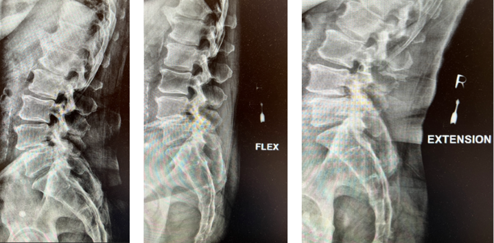
The grade 1 spondylolisthesis at L4/L5 requires both decompression at the L4/L5 level but also fusion because of the instability with motions. Here are three standing x-rays: one in the neutral position, one in flexion, and one in extensions. Notice the movement in the vertebral bodies at L4 and L5. This is abnormal motion and it can contribute to back pain and increase stenosis in certain positions. Thus, it requires decompression and stabilization with fusions.

Image 2. This shows the lumbar spine in motion. Notice the L4 and L5 level changes with flexion and extension. Notice the L2 and L3 levels do not have that type of motion. This is instability.

Image 3. Post-op x-rays that show L4/L5 transforaminal lumbar interbody fusion (TLIF). There was also removal the L2/L3 disc herniation but no need for instrumentation.
The herniated disc only requires decompression and removal of the herniated disc. Notice in the same flexion extension dynamic x-rays. There’s no abnormal motion at the L2/L3 level.
Dr. Imani was able to decompress both the L2/L3 level (with disc herniation removal) and the L4/L5 level. Additionally, L4/L5 level was fused using titanium screw and an expandable interbody cage. This was performed with a very small incision. The patient has no more leg pain and back pain, and is doing very well!
References
1. Austevoll IM, Hermansen E, Fagerland MW, et al. Decompression with or without Fusion in Degenerative Lumbar Spondylolisthesis. New England Journal of Medicine 2021; 385: 526 – 538.
2. Kreiner DS, Hwang SW, Easa JE, et al. An evidence-based clinical guideline for the diagnosis and treatment of lumbar disc herniation with radiculopathy. Spine J. 2014 Jan; 14(1): 180 – 91.
CATEGORY: SPINE//TREATMENT OF SPONDYLOLISTHESIS AND DISC HERNIATION
Treatment of Spondylolisthesis and Disc Herniation